
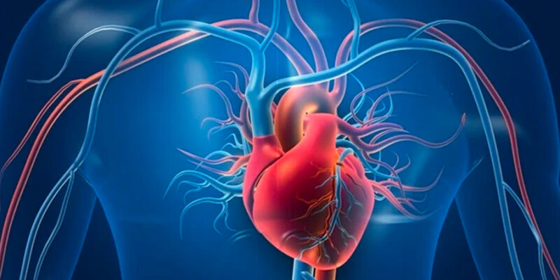
The heart is a complex muscular structure that is the central organ of the circulatory system. It is a four-chambered organ that functions primarily to pump blood throughout our bodies. It is as big as the fist of a human hand. It is located between the lungs, more on the left side, and rests on the diaphragm.
There are four chambers in a heart namely atrium and ventricle, two each. The structure of heart can be well understood by studying these four chambers that are described below.
Chambers of the Heart
1. Right Atrium
The right atrium is a smooth-walled chamber that receives deoxygenated blood from several sources. The superior and inferior vena cava are two major vessels that open into this chamber. They bring back the deoxygenated blood from systemic circulation. Another source is the coronary sinus which brings deoxygenated blood from all the coronary arteries.
2. Right Ventricle
The right ventricle is a muscular chamber that receives deoxygenated blood from the right atrium by contraction. The right ventricle contracts to force the blood to the lungs through the pulmonary trunk for oxygenation of blood.
3. Left Atrium
The left atrium is situated just behind the right atrium and forms the majority of the base in the heart. Just like the right atrium, it is also smooth-walled. The pulmonary veins open into the left atrium and bring freshly oxygenated blood from the lungs into the heart.
4. Left Ventricle
The left ventricle is the main powerhouse of the heart that transports oxygenated blood to the systemic circulation. The left ventricle is almost three times thicker and bigger. It forms the apex of the heart. The aorta opens from the left ventricle that carries blood to the whole body through smaller arteries.
Thus, the vena cava, the pulmonary artery, the pulmonary vein, and the aorta make up the major blood vessels of the heart. They are also referred to as the great vessels.
Layers of the Heart
The heart is enclosed in a pericardial sac that is filled with pericardial fluid. The pericardial fluid absorbs shocks and protects the heart from external trauma if any. There are three layers covering the heart:
- Epicardium: It is the innermost layer of the pericardium that forms the outer covering of the heart.
- Myocardium: The myocardium is the thick and muscular middle layer of the heart.
- Endocardium: The endocardium is the innermost layer of the heart that lines the four chambers.
The whole phenomenon of contraction and relaxation of the heart muscles to produce one heartbeat is called the cardiac cycle. The relaxation of the heart when it fills up with blood is called diastole and the contraction of heart that leads to the pumping of blood is known as systole. The four chambers and the valves of the heart function together to pump blood throughout the body.
Valves of the Heart
There are four valves in the heart. Let us discuss each of them separately.
1. Tricuspid Valve
The tricuspid valve is an atrioventricular valve located between the right atrium and right ventricle. As the name suggests, it is made up of three flaps. The right atrium contracts to pass the deoxygenated blood from the right atrium to the right ventricle. The tricuspid valve prevents the backflow of blood back into the right atrium.
2. Pulmonary Valve
The pulmonary valve is a semilunar valve that is situated between the right ventricle and pulmonary trunk. The pulmonary valve prevents the regurgitation of the deoxygenated blood from the pulmonary trunk back to the right ventricle. It is also made up of three cusps.
3. Bicuspid Valve
The bicuspid valve is another atrioventricular valve that is located between the left atrium and left ventricle. It is made up of two cusps. The bicuspid valve, also known as the mitral valve, facilitates the transfer of oxygenated blood from the left atrium into the left ventricle and prevents blood flow.
4. Aortic Valve
The aortic valve is another semilunar valve present between the left ventricle and aorta. It is made up of three cusps. It allows the passage of oxygenated blood from the left ventricle to the aorta and subsequently to the whole body.
Visit BYJU’S Biology to learn more in detail about the human heart. Students can also learn more by watching different subject related YouTube videos by subscribing to YouTube videos.


Comments